Entomology and
Applied Science Letters
Applied Science Letters
 2023
Volume 10
Issue 3
2023
Volume 10
Issue 3

Synovial joints are an important part of the body and their lubrication plays a crucial role in joint movement. This study was conducted to investigate the surface ultrastructure of synovial fluid in indigenous cattle to identify its diversity from other species to improve the diagnosis and prognosis of joint diseases in this species. The present study was performed in the central region (state Madhya Pradesh) of India. Synovial fluid was collected from the knee joint of the young calves (group I-six months to one year) and adults of indigenous cattle (group II-4-5 years). The study was carried out with the help of scanning electron microscopy (SEM). The results showed cross-shaped intertangled stringy networks and extracellular vesicles in the synovial fluid of the cattle. The presence of both structures supports the presence of boundary and hydrodynamic lubrication, however, the abundant presence of cross stringy network and unilamellar vesicles suggest that boundary lubrication might be a prime mode of lubrication.
INTRODUCTION
Synovial joints are an important topic of research in human beings as they suffer from various arthritis conditions that affect their mobility. But nowadays due to changes in the rearing system, as intensive housing is the choice of rearing due to scarcity of pasture lands and increased urbanization, animals also suffer from joint-related ailments at an early age which affects their life span as well as productivity [1, 2].
Synovial joints are an important part of the body and synovial joint lubrication is the best example of lubrication depending on the nature of articulating surfaces and synovial fluid. Many experimental and theoretical studies have attempted to understand the powerful mechanisms of lubrication of healthy synovial fluid (SF) and their involvement in joint failure [3]. However, the mechanisms involved are not completely known [4].
Different theories of lubrication were proposed out of which boundary lubrication and hydrostatic theories are important to explain how the lubrication takes place between these components of synovial joints [5]. They stated that boundary lubrication mitigates stick slips so it is important for steady motion and heavy load application while hydrodynamic fluid film lubrication occurs with pressurized motion and deformation acting to drive the viscous lubricant through the gap between two surfaces.
Attempts to describe a single mode of lubrication to synovial joints have undoubtedly delayed the emergence of a satisfactory overall picture of the performance of nature’s bearing so there is a possibility of a multimode of lubrication in a single species [6]. Synovial fluid is a pseudo-plastic (shear-thinning) fluid, both in normal and pathological conditions. The fluid exhibits elasticity and a normal-force effect [7]. The low friction in joints is associated with a full film of lubricant which separates the surfaces and high friction results formation of thin films of localized boundary friction due to asperity contacts [8].
Chemically It was supposed that the viscosity of the hyaluronic acid plays an important role in the lubrication ability of the synovial fluid, however, Tadmor et al. [9] stated that hyaluronic alone does not provide any exceptionally low friction force. This is the association of phospholipid with hyaluronic acid which results in the preparation of a potent lubricating agent [10]. The ability of biochemical compounds to lubricate surfaces relies not only on their concentration but also on their ultrastructural interactions so detailed knowledge about the ultrastructure of synovial fluid is of utmost importance. The three-dimensional surface morphology of the synovial fluid can easily be explored with the help of scanning electron microscopy [11].
Despite the development of different techniques of physicochemical characterization of synovial fluid definite ultrastructural interaction in different species is not very well known. In a few species presence of multilamellar vesicles and gel in type structure is reported [12, 13] however variability in this structural pattern with the change in species is under investigation.
Looking into the importance of the synovial fluid because of its structural and physiological nature the present study aimed to explore the surface ultrastructure of synovial fluid in indigenous cattle to identify the variability from other species to improve the diagnosis and prognosis of joint diseases in this species.
MATERIALS AND METHODS
The present study was performed in the central region (state Madhya Pradesh) of India. Synovial fluid was collected from the knee joint of young calves (group I-six months to one year) and adult (group II-4-5 years) indigenous cattle. 200 µl synovial fluid was preserved in 500 µl of 2.5% glutaraldehyde solution in 0.05 M phosphate buffer at 4 °C for 6-8 hrs. The preserved fluid was centrifuged at 8000 rpm for 10 minutes after that two batches of samples were prepared. In one batch sample was poured on 0.22 µm millipore filter paper and in another batch sample was placed over the cover slip diluted with the distilled water. Both batches were air dried for two days and then coating with gold particles was performed. Observations were made with the Zeiss Evo 18 Scanning Electron Microscope at SAIF, AIIMS, Delhi. Ethical permission for the experiment was granted by the institutional ethical committee.
RESULTS AND DISCUSSION
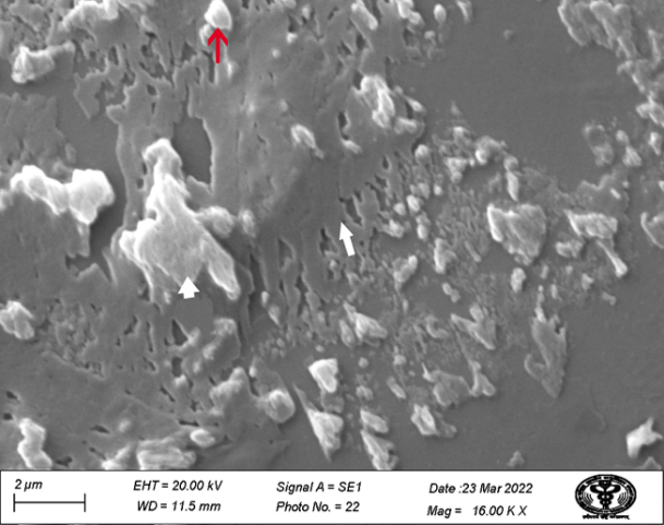
Synovial fluid's biochemical components manifested themselves in the current research as cross-shaped intertangled stringy networks and extracellular vesicles. The cross-shaped stringy network may have been created by the complicated lipid-protein and hyaluronic acid complex tangling. A closer look at the network revealed that it was composed of tiny aggregates with ambiguous morphology. It represents the bulk portion of the synovial fluid (Figure 1).
|
|
|
Figure 1. Scanning electron micrograph of knee joint synovial fluid (group II, coverslip) showing the cross-shaped stringy network of an acid protein complex (arrow), synoviocyte B (arrowhead), and irregularly shaped cells ( |
The presence of the same structure in the synovial fluid of humans was also reported by Walker et al. [14]. The same finding was supported by the Seller et al. [8]. Pasquali-Ronchetti et al. [15] also studied the interaction of different molecular weight hyaluronic acid and phospholipid vesicles. In the study, they attributed that the high molecular weight of hyaluronic acid is responsible for the formation of sheet-like structures. They further mentioned that aggregation is further influenced by the temperature as high temperature may affect the melting procedure of the lipid vesicles. So, the presence of a significant sheet-like structure might be due to the tropical nature of the Country.
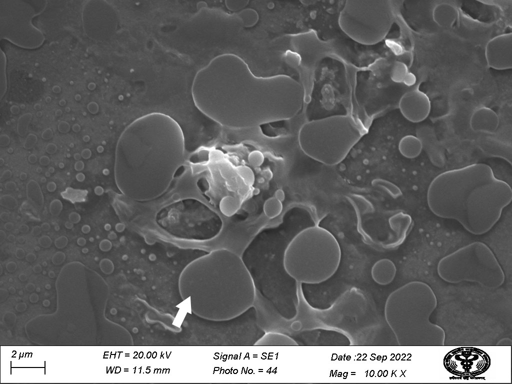
The second extracellular vesicle of bovine species had a lamellated appearance and a grey center encircled by an electron-lucent periphery. Usually unilamellar structure was present however oligolamellar appearance was also observed scatterdly (Figure 2).
|
|
|
Figure 2. Scanning electron micrograph of knee joint synovial fluid (group II, coverslip) showing electron-dense layered vesicles (arrowhead). |
Allen and Drauglis [16] detailed and reviewed boundary lubrication and stated that mono-lamellar film is most suitable for boundary lubrication due to adsorption on the surface, however, the multilamellar film can also stand with high shear forces due to high resistance against the force. Variation between species is attributed to the type of motion as well as the application of load.
The extracellular vesicles a messenger elements carrying various compounds and can be used for the early detection of different arthritic conditions [17]. Matei et al. [12] and Ben-Trad et al. [13] described the presence of multilamellar vesicles in the synovial fluid of species equine humans, rat on the other hand in humans and dogs, respectively. Pasquali-Ronchetti et al. [15] stated that in the presence of low molecular weight hyaluronic acid, phospholipid moiety remains in the vesicular form so it might be possible that in healthy animals molecular weight of hyaluronic acid remains variable, and found in proportion and provide stability to lubrication [15]. During different ailments might be this proportion misbalanced. In the present study difference in the electron density at the core and periphery of vessels was further supported by the study of Sava et al. [4]. They evaluated artificial and natural synovial fluid and found that natural synovial fluid has gel in a gel-in-type arrangement where hyaluronic acid and protein components form the core and phospholipids form the periphery.
CONCLUSION
The presence of both cross-stringy network and vesicular structure in the synovial fluid of the cattle supports the presence of boundary lubrication and hydrodynamic lubrication. The presence of a significant amount of cross-stringy network in the specimen indicates that the boundary lubrication is prime mode because of the heavy body weight and slow steady motion. The process of dehydration and the effect of temperature in tropical areas can't be denied. Further study is required to see the effect of higher body weight and temperature in different climate zones to formulate remedies for arthritis.
ACKNOWLEDGMENTS: We are thankful to Sophisticated Analytical Instrumentation Facility AIIMS, New Delhi, India for providing help, instrumental facility, and guidance during the research.
CONFLICT OF INTEREST: None
FINANCIAL SUPPORT: None
ETHICS STATEMENT: Ethical certificate no: 54/IAEC/vety/2020 date: 26/10/2020.