Creative Commons License 2023 Volume 10 Issue 2

Development of a Method for Studying the Structural Organization of Bone Tissue Using Computed Tomography


, , , , ,
Abstract

This scientific work describes a new way of analyzing the organization of bone tissue using computed tomography(CT). The information obtained as a result of such a study can be used to diagnose bone diseases, as well as in research in the field of biology and medicine. The common lizard was chosen as a test subject, and the bones of the proximal tail vertebrae were selected as samples for the study of the structural organization of bone tissue. The samples must be clean and in good condition so that the data obtained are reliable. In this study, a laboratory micro tomograph was utilized. In this study, we investigated both the spatial distribution of the X-ray absorption coefficients and the distribution of the elemental compositions within the bone tissue which have been largely unstudied so far. The absorption densities of these vertebrae have been found to vary greatly depending on the experimental conditions and pharmaceutical manufacturing processes. Areas of increased density located closer to the central canal of the vertebra were identified.


How to cite this article
Vancouver
Dzhatdoeva KH, Adzhiev RS, Bidzhieva MU, Tsoraev HM, Klishchenko RV, Petinova YA. Development of a Method for Studying the Structural Organization of Bone Tissue Using Computed Tomography. Entomol Appl Sci Lett. 2023;10(2):19-24. https://doi.org/10.51847/uoTObx18IP
APA
Dzhatdoeva, K. H., Adzhiev, R. S., Bidzhieva, M. U., Tsoraev, H. M., Klishchenko, R. V., & Petinova, Y. A. (2023). Development of a Method for Studying the Structural Organization of Bone Tissue Using Computed Tomography. Entomology and Applied Science Letters, 10(2), 19-24. https://doi.org/10.51847/uoTObx18IP
Downloads: 43
Views: 182

Development of a Method for Studying the Structural Organization of Bone Tissue Using Computed Tomography


Kamila Hamitovna Dzhatdoeva1, Rustam Salimovich Adzhiev2*, Madina Umarovna Bidzhieva1, Harun Muratovich Tsoraev3, Rostislav Valerievich Klishchenko4, Yulia Alexandrovna Petinova1


1Department of Therapy, Faculty of Medicine of Stavropol State Medical University, Stavropol, Russia.

2Department of Radiology, Medical Faculty of Stavropol State Medical University, Stavropol, Russia.

3Stavropol Regional Clinical Oncological Dispensary, Stavropol, Russia.

4Department of Therapy, Institute "Medical Academy named after S.I. Georgievsky", Crimean Federal University named after V.I. Vernadsky", Simferopol, Russia.


ABSTRACT

This scientific work describes a new way of analyzing the organization of bone tissue using computed tomography(CT). The information obtained as a result of such a study can be used to diagnose bone diseases, as well as in research in the field of biology and medicine. The common lizard was chosen as a test subject, and the bones of the proximal tail vertebrae were selected as samples for the study of the structural organization of bone tissue. The samples must be clean and in good condition so that the data obtained are reliable. In this study, a laboratory micro tomograph was utilized. In this study, we investigated both the spatial distribution of the X-ray absorption coefficients and the distribution of the elemental compositions within the bone tissue which have been largely unstudied so far. The absorption densities of these vertebrae have been found to vary greatly depending on the experimental conditions and pharmaceutical manufacturing processes. Areas of increased density located closer to the central canal of the vertebra were identified.

Keywords: Computed tomography, Bone, Cartilage, X-ray introscopy.


INTRODUCTION


Advances in the fields of nano- and micro technologies, microbiology, and biomedical diagnostics, are associated with the development of modern methods for non-destructive studies of the internal structure of complex objects [1-3]. One of the currently existing methods of such diagnosis is the method of X-ray introscopy [4]. X-ray computed tomography techniques are advanced methods of X-ray introscopy or, more specifically, X-ray microscopy and are excellent for a wide range of investigations [5-7].

Wilhelm Conrad Roentgen discovered X-ray radiation [8]. He was the first person to publish his paper on X-rays. Due to the high penetrating power of X-ray radiation, non-destructive testing of the studied objects became possible for researchers [9]. X-ray computed tomography has become relevant in many fields of science, such as medicine, geology, materials science, archaeology, food science, etc. [10-14].

Many scientific groups around the world are currently working on the development of X-ray microscopy [15, 16]. After the creation of synchrotron radiation sources in the middle of the last century, many experiments have been carried out with them [17, 18]. Modern microelectronics developments have created digital detectors with high spatial resolution making it possible to acquire images within seconds [19, 20]. The latest developments in the fields of materials science, microelectronics, and nanotechnology contribute to the development of manufacturing techniques for the production of X-ray optics. This improves the quality, significantly increases the information content of the images obtained, and increases their spatial resolution [21].

Microtomographs are an important tool for studying biological samples at the microscopic level [22]. Some of the applications of microtomography in biology include:

  1. Study of bone structure: Microtomographs allow to study of the structure of bone tissue at the microscopic level. This can help in the study of various bone diseases, such as osteoporosis [23].
  2. Study of cell structure: Microtomographs allow you to study the structure of cells at the nanometer level. This can help in the study of various biological processes, such as cell division or interaction with the environment [24].
  3. Study of the structure of small organisms: Microtomographs allow you to study the structure of small organisms, such as bacteria and algae [25]. This can help in the study of their functions and role in ecosystems.
  4. Study of the structure of materials: Microtomographs can be used to study the structure of various materials, such as ceramics, polymers, and composites [26]. This can help in the development of new materials and improve their properties.
  5. Study of the structure of samples and artifacts: Microtomographs can be used to study the structure of various samples and artifacts, such as archival documents, archaeological finds, and technical details [27]. This can help in their preservation and reconstruction.

In this work, the internal structure of vertebrate bone tissue was investigated. Of interest is both the spatial distribution of the X-ray absorption coefficient and the distribution of the elemental composition within the bone tissue, since these issues have been little studied to date.

MATERIALS AND METHODS

The common lizard (Lacerta agilis) was chosen as the subject of the study. The common lizard (Lacerta agilis) was chosen as the object of the study. The agile lizard, or common lizard, is a type of lizard in the real lizard family. It is a scaly reptile that is up to 25 cm long, with relatively short limbs and a tail that is almost twice its trunk in length. It is brown with dark and light spots and stripes. It has a light lower abdomen. The skull is flexibly connected to the spine but has only one appendage. The skeleton of a lizard is notable for its well-developed ribs from its chest.  Its dry skin is covered with horny scales as shields [28]. The divisions of the reptilian digestive system include the oral cavity, pharynx, stomach, digestive glands, pancreas, liver, small and large intestines, and cloaca. The excretory organs include the kidneys, ureters, and bladder. The skeleton is entirely bony. The spine is separated into five sections: cervical, thoracic, lumbar, sacral, and caudal. The head is mobile because of the elongation of the neck and the presence of two specialized cervical vertebrae [29].

The cervical spine has several vertebrae and the first two of which allow the head to turn in any direction. The thorax secures the shoulder girdle through the chest and supports the front legs. The lumbar region provides torso flexion that aids movement. The powerful sacral region already consists of two vertebrae and the girdle of the hind limbs is paralyzed. The long tail section provides balancing movements of the tail [30]. Breathing is only through the lungs. A suction breathing mechanism that is more perfect than in amphibians (respiration occurs by changing the volume of the chest). Conductive airways (larynx, trachea, bronchi) are well developed. The lining and septum of the lungs have a cellular structure. The heart has three chambers and consists of one ventricle and two atria.

An incomplete septum is formed in the ventricle. Although the macrocirculatory and microcirculatory circuits are not completely separated, the venous and arterial flows are more restricted, allowing the reptile body to be supplied with more oxygenated blood. Venous blood from all organs of the body enters the right atrium, and arterial blood from the lungs enters the left atrium. When the ventricle contracts, its incomplete septum reaches the dorsal wall and separates the right and left halves. From the left half of the ventricle, arterial blood enters the vessels of the brain and the anterior part of the body, from the right half venous blood, goes to the pulmonary artery and then to the lungs. The trunk receives mixed blood from both halves of the ventricle.

The brain is more developed, especially the hemispheres of the forebrain (responsible for complex instincts), the visual lobes, and the cerebellum (coordinator of movements). The senses are more complicated. Reptile eyes distinguish between mobile and stationary objects. The lens in the eyes can not only move but also change its curvature. Lizards have movable eyelids. In the organs of smell, part of the nasopharyngeal passage is divided into the olfactory and respiratory departments. The internal nostrils open closer to the pharynx, so reptiles can breathe freely when they have food in their mouth [31].

To check the structural organization of the lizard's bone tissue, the bones of the proximal tail vertebrae were selected as samples for the study. Two samples were examined, and the characteristic dimensions of these objects did not exceed several millimeters.

Bone tissue sampling is an important step in microtomographic examination. It determines whether researchers come to accurate and complete conclusions:

  1. Bone tissue should be presented in its natural state, without any changes, for example, intact.
  2. The bone tissue sample should be presented in sizes not exceeding the overall capabilities of the microtomograph.
  3. The optimal sample size should be at least 1 mm in diameter and at least 3 mm in length.
  4. The optimal number of samples to get an accurate result should be at least two samples.

A sample of bone tissue must be taken from a place that has not changed its structure and has not narrowed as a result of mechanical action.

It is prohibited to use a sample that has been treated with a chemical substance for preservation.

The bone tissue sample must be preserved under special conditions to prevent deformation that affects the result.

Tomographic studies of the volumetric structure of animal bone tissue samples were carried out on a laboratory X-ray microtomograph HeliScan micro CT (Thermo Scientific) (FEI, USA) in two X-ray modes (5.4 and 12.0 kV).

RESULTS AND DISCUSSION

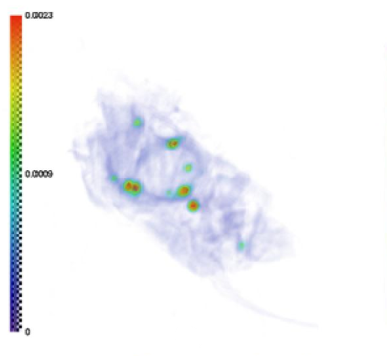
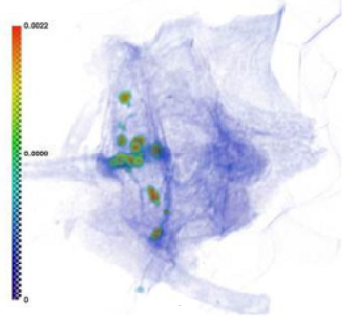
Tomographic studies of the volumetric structure of animal bone tissue samples were performed on a laboratory X-ray microtomograph. Typical results of the restoration of the internal structure of the samples are shown in Figure 1.


a)

b)

Figure 1. Tomographic reconstruction of the vertebrae of a lizard samples a, b


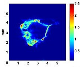
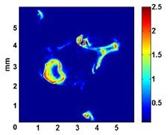
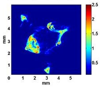
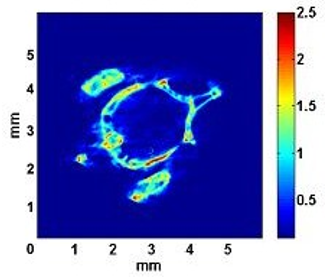
The studies were carried out at various X-ray energies (5.4 and 12.0 kv). In the samples, areas that absorb significantly more than the rest of the bone tissue were found (Figure 2). This may indicate that it is in these areas (located closer to the central canal of the vertebra) that heavy elements are localized. In addition, the experimental results show that the peripheral areas of the studied samples do not contain elements with an atomic number greater than 20 (which corresponds to calcium atoms) or their concentration is less than the sensitivity of the method, since the absorption of X-ray radiation here varies in proportion to its wavelength.


a)

b)

c)

d)

Figure 2. Cross-sections of the spine of a lizard with a nimble

It was found that, depending on the experimental conditions and methods of preparation of drugs, the absorbing density of these vertebrae can vary markedly. Areas of increased density located closer to the central canal of the vertebra were identified. It should be noted that the ratio of absorption in different areas of bone tissue changes with varying wavelengths of probing X-ray radiation. This indirectly indicates the uneven distribution of the elemental composition in the volume of bones. The correlation of X-ray fluorescence analysis data with the results of X-ray microtomography has been established.

According to X-ray fluorescence analysis, the presence of several heavy elements (Fe, Ni, Cu, Zn, Br, Sr) in these structures was established for the first time and their uneven distribution was revealed. A comparison of electron microscopy data and X-ray fluorescence analysis shows that these elements are located in the depth of the bone tissue (clearly deeper than at a distance of 10 microns from its surface), which also corresponds to the results of X-ray microtomography.

CONCLUSION

In this work, the internal structure of vertebrate bone tissue was investigated. Of interest is both the spatial distribution of the X-ray absorption coefficient and the distribution of the elemental composition within the bone tissue.  During computer microtomography, areas were found in the samples that absorb significantly more than the rest of the bone tissue. This suggests that heavy elements (Fe, Ni, Cu, Zn, Br, Sr) are localized in those areas. Thus, the uneven distribution of the elemental composition in the volume of bones is revealed.

ACKNOWLEDGMENTS: The authors are thankful to colleagues from Dagestan State Medical University for assistance in experiments.

CONFLICT OF INTEREST: None

FINANCIAL SUPPORT: None

ETHICS STATEMENT: The protocol for experiments with beetles complied with the requirements of the European Convention for the Protection of Vertebrate Animals used for Experimental and other Scientific Purposes.


References

1.       Zhao DD, Liu XE, Yang SM, Yu S, Tian GL, Ma JF, et al. Study on Non-Destructive Testing of Guqin Interior Structure Based on Computed Tomography. Guang Pu Xue Yu Guang Pu Fen Xi. 2015;35(12):3519-23. Chinese.

2.       Shevchenko YS, Plohova DP, Bulakhova IN, Mishvelov AE, Kubalova ME, Badriev GB. Experience in carrying out magnetic resonance imaging with the use of specialized protocols and programs computer post-processing. Pharmacophore. 2020;11(2):77-81.

3.       Mezhidov BS, Belyaeva AA, Kh SM, Bimarzaev S, Bektashev A, Shekhshebekova AM, et al. Prospects for creating 3D models of internal organs based on computer and magnetic resonance imaging images in emergency surgery and resuscitation. Pharmacophore. 2021;11(1):8-14. doi:10.51847/3TLcii4n42

4.       Du Plessis A, Yadroitsev I, Yadroitsava I, Le Roux SG. X-ray microcomputed tomography in additive manufacturing: a review of the current technology and applications. 3D Print Addit Manuf. 2018;5(3):227-47. doi:10.1089/3dp.2018.0060

5.       Curto M, Kao AP, Keeble W, Tozzi G, Barber AH. X-ray computed tomography evaluations of additive manufactured multi-material composites. J Microsc. 2022;285(3):131-43. doi:10.1111/jmi.13034

6.       Guntoro PI, Ghorbani Y, Koch PH, Rosenkranz J. X-ray Micro-computed Tomography (µCT) for Mineral Characterization: A Review of Data Analysis Methods. Minerals. 2019;9(3):183. doi:10.3390/min9030183

7.       Fogelqvist E, Kördel M, Carannante V, Önfelt B, Hertz HM. Laboratory cryo x-ray microscopy for 3D cell imaging. Sci Rep. 2017;7(1):1-8. doi:10.1038/s41598-017-13538-2

8.       Franke OC. Wilhelm Conrad Roentgen and other X-ray pioneers. Bull Hist Dent. 1983;31(1):11-7.

9.       Jee KW, Zhang R, Bentefour EH, Doolan PJ, Cascio E, Sharp G, et al. Investigation of time-resolved proton radiography using an x-ray flat-panel imaging system. Phys Med Biol. 2017;62(5):1905-19. doi:10.1088/1361-6560/aa5a43

10.   Schendrigin IN, Timchenko LD, Rzhepakovsky IV, Avanesyan SS, Sizonenko MN, Grimm WD, et al. Clinical and pathogenetic significance of amylase level and microtomographic index of synovial fluid in various joint lesions. Sovrem Tehnol Med. 2022;14(6):42. doi:10.17691/stm2022.14.6.05

11.   Abrosimov KN, Gerke KM, Fomin DS. Tomography in Soil Science: From the First Experiments to Modern Methods (A Review). Eurasian Soil Sci. 2021;54:1385-99. doi:10.1134/S1064229321090027

12.   Abrosimov KN, Gerke KM, Semenkov IN. Otsu’s Algorithm in the Segmentation of Pore Space in Soils Based on Tomographic Data. Eurasian Soil Sci. 2021;54(4):560-71. doi:10.1134/S1064229321040037

13.   Nagdalian AA, Rzhepakovsky IV, Siddiqui SA, Pskov SI, Oboturova NP, Timchenko LD, et al. Analysis of the content of mechanically separated poultry meat in sausage using computing microtomography. J Food Compost Anal. 2021;100:103918. doi:10.1016/j.jfca.2021.103918

14.   Iammarino M, Miedico O, Petrella A, Mangiacotti M, Chiaravalle AE. Innovative approaches for identifying a mechanically separated meat: evaluation of radiostrontium levels and development of a new tool of investigation. J Food Sci Technol. 2020;57(2):484-94. doi:10.1007/s13197-019-04076-y

15.   Remizova AA, Bitarov PA, Epkhiev AA, Remizov NO. Reparative-regenerative features of bone tissue in experimental animals treated with titanium implants. J Adv Pharm Educ Res. 2022;12(2):110-6.

16.   Alshammari E. Routine prescription of calcium in immobile females receiving medroxyprogesterone to prevent osteoporosis/bone loss. J Adv Pharm Educ Res. 2020;10(3):155-9.

17.   Alanazi AM, Alotaibi HD, Alahmari SA, Almutairi AK, Babakr SA, Abdrabalnabi HA, et al. Hip Bone Fracture Diagnosis and Management. Arch Pharm Pract. 2019;10(4):29-32.

18.   Mostafavi Z, Houshyar J, Aliasgarzadeh A, Aghamohammadzadeh N, Sadra V, Khani M, et al. Comparison of the Bone Mineral Density between Acromegaly Patients and Healthy Individuals. Arch Pharm Pract. 2019;10(4):15-21.

19.   Bouckaert C, Vandenberghe S, Van Holen R. Evaluation of a compact, high-resolution SPECT detector based on digital silicon photomultipliers. Phys Med Biol. 2014;59(23):7521-39. doi:10.1088/0031-9155/59/23/7521

20.   Hussein K. Ethnic Differences in Bone Mass and Vitamin D status in Young Women Living in the UK. Int J Pharm Res Allied Sci. 2022;11(3):98-107.

21.   Yoshida S, Sato E, Oda Y, Yoshioka K, Moriyama H, Watanabe M. Triple-sensitivity high-spatial-resolution X-ray computed tomography using a cadmium-telluride detector and its beam-hardening effect. Appl Radiat Isot. 2020;159:109089. doi:10.1016/j.apradiso.2020.109089

22.   Cercos-Pita JL, Fardin L, Leclerc H, Maury B, Perchiazzi G, Bravin A, et al. Lung tissue biomechanics imaged with synchrotron phase contrast microtomography in live rats. Sci Rep. 2022;12(1):5056. doi:10.1038/s41598-022-09052-9

23.   Sarmento VA, de Oliveira Gonzalez TFL, Lopes RT, Novaes M, Borges SM, Rubira-Bullen IRF. A Comparative Study of Multidetector Computed Tomography, Cone Beam Computed Tomography, and Computed Microtomography on Trabecular Bone Structures in the Human Mandible: An Ex Vivo Study. J Comput Assist Tomogr. 2021;45(4):552-6. doi:10.1097/RCT.0000000000001191

24.   Tamminen I, Lehto K, Hannula M, Ojansivu M, Johansson L, Kellomäki M, et al. A tube-source X-ray microtomography approach for quantitative 3D microscopy of optically challenging cell-cultured samples. Commun Biol. 2020;3(1):548. doi:10.1038/s42003-020-01273-w.

25.   Shearer T, Bradley RS, Hidalgo-Bastida LA, Sherratt MJ, Cartmell SH. Three-dimensional visualization of soft biological structures by X-ray computed micro-tomography. J Cell Sci. 2016;129(13):2483-92. doi:10.1242/jcs.179077

26.   da Silva ÍB. X-ray Computed Microtomography technique applied for cementitious materials: A review. Micron. 2018;107:1-8. doi:10.1016/j.micron.2018.01.006

27.   Phan J, Ruspini LC, Lindseth F. Automatic segmentation tool for 3D digital rocks by deep learning. Sci Rep. 2021;11(1):19123. doi:10.1038/s41598-021-98697-z

28.   Latip MQA, Tengku Azizan TRP, Ahmad H, Abu Hassim H, Noor MHM, Mikail M. Blood Profiling of Captive and Semi-Wild False Gharial in Peninsular Malaysia. Animals (Basel). 2021;11(6):1481. doi:10.3390/ani11061481

29.   Freitas ETF, Moreira AMS, de Paula RS. Ultrastructure of the gill ciliary epithelium of Limnoperna fortunei (Dunker 1857), the invasive golden mussel. BMC Zool. 2020;7(1):6. doi:10.1186/s40850-022-00107-y

30.   Horváthová T, Baláž M, Jandzik D. Reproduction and morphology of the common lizard (Zootoca vivipara) from montane populations in Slovakia. Zoolog Sci. 2013;30(2):92-8. doi:10.2108/zsj.30.92

31.   Atencia PL, Castillo CJ, Montes LF. Use of microhabitat and activity patterns of two lizard species from a seasonal dry forest in northern Colombia. Neotrop Biol Conserv. 2020;15(2):153-64. doi:10.3897/neotropical.15.e49713


Entomology and Applied Science Letters is an international double-blind peer reviewed publication which publishes scientific research & review articles related to insects that contain information of interest to a wider audience, e.g. papers bearing on the theoretical, genetic, agricultural, medical and biodiversity issues. Emphasis is also placed on the selection of comprehensive, revisionary or integrated systematics studies of broader biological or zoogeographical relevance. In addition to full-length research articles and reviews, the journal publishes interpretive articles in a Forum section, Short Communications, and Letters to the Editor. The journal publishes reports on all phases of medical entomology and medical acarology, including the systematics and biology of insects, acarines, and other arthropods of public health and veterinary significance.

Announcement and Advertisement
Announcements regarding scientific activities such as conferences, symposium, are published for free. Advertisements can be either published or placed on website as banners.
open access
Entomology and Applied Science Letters supports the submission of entomological papers that contain information of interest to a wider reader groups e. g. papers bearing on taxonomy, phylogeny, biodiversity, ecology, systematic, agriculture, morphology. The selection of comprehensive, revisionary or integrated systematics studies of broader biological or zoogeographical relevance is also important. Distinguished entomologists drawn from different parts of the world serve as honorary members of the Editorial Board. The journal encompasses all the varied aspects of entomological research.